Programme

Opening Ceremony

09:30 – 09:45

Welcome Remarks

Group Photo

Keynote Talks

09:45 – 10:20
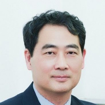
Abstract: Neural information processing in the brain is characterized by extensive interactions between feedforward and feedforward signals. It has been suggested that the feedforward-feedback recurrent processing is the basis for visual awareness. However, the functional significance of feedback signals remains unclear. In a series of studies, we investigated the nature and impact of feedback signals in visual object processing and visual adaptation. Results show that feedback signals play important roles in object recognition, are temporally flexible, task-dependent, and are more important than the feedforward signal in determining neuronal sensitivity in the visual cortex.

Bio: Prof. HE Sheng obtained his Ph.D in psychology from UC San Diego, followed by post-doctoral training at Harvard University. He was on the faculty in the psychology department at the University of Minnesota from 1997 till 2020. He is currently serving as the director of the State Key Laboratory of Brain and Cognitive Science at the Institute of Biophysics, Chinese Academy of Sciences. His main research interests are in human cognitive neuroscience, especially visual cognition. He uses psychophysical as well as brain imaging approaches to study mechanisms of visual object recognition, attention, and consciousness.

Prof. Zhen YUAN

10:20 – 10:55
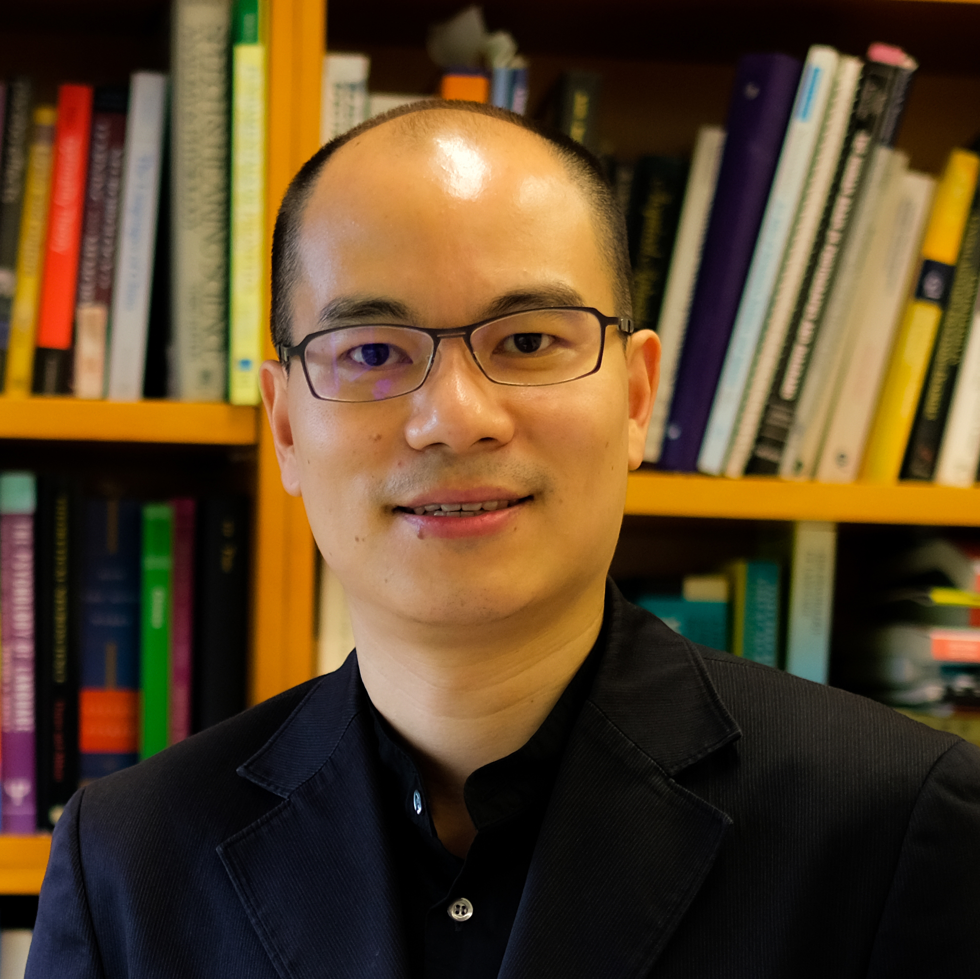
Abstract: Language neuroscience can make important contributions to the broader agenda of precision medicine by capitalizing on what we know about the neurobiology of language learning to improve the precision of detection and intervention of language-related neurodevelopmental conditions. In this presentation, I report findings from a series of studies in which we use neural data collected as early as infancy to construct predictive models to forecast language developmental outcomes at the individual child level. These studies include data from preterm and term-born infants, cochlear implant candidates, and children with a confirmed diagnosis or are at elevated likelihood of autism. Our results show that predictive models using neural data to forecast language developmental outcomes often outperform those that use standard clinical and demographic measures as predictors. In some cases, we have sufficient data to validate the models using unseen data or to evaluate the models’ generalization to cross-site and cross-language data. Besides their potential clinical applications, our neural predictive models also provide opportunities to address more basic questions about the neurobiology of language. For example, we ask whether cortical and subcortical development interacts with native and non-native speech processing in infancy, and whether this interaction forms the basis of spoken language development. In children who are hearing impaired, we examine whether brain regions that are resilient to reduced auditory/spoken language input are those that promote spoken language when hearing is facilitated by cochlear implantation. In both typical and atypical populations, we are in the process of testing whether individual-child predictions can inform the design and prescription of different types of early intervention and enhancement strategies so that language development can be optimized for all children.

Bio: Prof. Patrick C. M. Wong holds the Stanley Ho Chair in Cognitive Neuroscience, is Professor of Linguistics, Otolaryngology and Psychology, and serves as the Founding Director of the Brain and Mind Institute at The Chinese University of Hong Kong (CUHK). Wong’s research covers a wide range of basic and translational issues concerning the neural basis and disorders of language and music. In 2021, he was named a Guggenheim Fellow for Humanities. Wong’s research has received public attention from media outlets such as The New York Times. He actively seeks to translate his research into clinical and educational solutions. One of his patented inventions, Precision Listening®, was awarded the Gold Medal with Congratulations of the Jury at the 2023 International Exhibition of Inventions Geneva. Wong has been Associate Vice-President (Research) at CUHK since 2023.

Prof. Zhen YUAN

10:55 – 11:15

Refreshment & Poster Session

Guest Talks

11:15 – 11:38
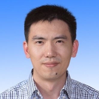
Abstract: Humans are remarkably adept at recognizing the motion of biological entities in complex visual scenes. Biological motion can be captured with a handful of point lights attached to the head and major joints of the body, and can be further decomposed into two components: global configuration and local motion. Whereas most previous studies have emphasized the contribution of global form to biological motion perception, our recent work indicates that local motion carries unique biological properties that can be processed independent of global configuration. These findings suggest that biological motion perception is a multilevel course encompassing the processing of both global configuration and local motion.
Bio: Prof. Yi Jiang currently holds the positions of Principal Investigator (PI) and Deputy Director at both the State Key Laboratory of Brain and Cognitive Science and the Institute of Psychology, Chinese Academy of Sciences. Additionally, he maintains an affiliation with the Department of Psychology at the University of Chinese Academy of Sciences. His research primarily focuses on exploring the neural underpinnings of human visual perception, attention, and awareness. To accomplish this, he employs a combination of psychophysics and brain imaging techniques.

Prof. Kuzma STRELNIKOV

11:38 – 12:01

Abstract: Structural magnetic resonance imaging (MRI) has become a critical tool in diagnosing age-related neurodegenerative diseases, with the potential to use MRI-informed metrices to revolutionise the field. Brain age matrices, including estimated brain age and brain-predicted age difference (brain-PAD), are computational indicators for measuring brain atrophy at individual level. An “older” brain age (i.e., positive score of brain-PAD) may indicate accelerated ageing. However, to effectively implement the computational matrices presents several barriers in real-world clinical populations. Therefore, in this presentation, we would have two aims: 1) investigate the MRI-informed brain age matrices in mild cognitive impairments (MCI) converters and their values in classifying MCI conversion (Study 1); 2) to examine whether individual’s brain age matrices can predict the responders to repetitive transcranial magnetic stimulation (rTMS) (Study 2). The pre-trained brain age model was constructed using MRI data from the Cam-CAN project (N=609). We found that greater brain age in MCI converters highlight the imaging signatures that may aid in classifying MCI converters at early stage. The elderly patients with younger brain age appears to be associated with better treatment responses to active rTMS.
Bio: Prof. Lu works as Assistant Professor and Deputy Director of Neuromodulation Clinic at the Department of Psychiatry, The Chinese University of Hong Kong. Her research interests focus on the development of personalized transcranial neuromodulation and using this technique to decode cognitive function and treatment brain disorders. Her studies have been published on renowned journals, such as JNNP, dialogues in clinical neuroscience, human brain mapping etc. Projects from her team have wined the best paper award on the Asian Oceanian Congress of Neurology and the Healthy Longevity Catalyst Award.

Prof. Kuzma STRELNIKOV

12:01 – 12:24

Abstract: Understanding how the human brain develops from childhood to adolescence, and identifying key systems that are linked to behavioral change, is a major goal in population neuroscience. This presentation recalls a short history of developmental population neuroscience and shows that the ventral attention network plays a crucial role in brain development and cognitive ability, with findings confirmed in cross-culture longitudinal cohorts.

Bio: Prof. Xi-Nian Zuo is the Fellow of the Organization of Human Brain Mapping, and currently serves as associate editors for Chinese Science Bulletin, Science Bulletin, China Scientific Data, Nature Scientific Data. He proposed Developmental Population Neuroscience and has focused on its cross-disciplinary discovery. Dr. Zuo has led multiple large-scale data-sharing projects including the Consortium for Reliability and Reproducibility (CoRR), 3R-BRAIN and Chinese Color Nest Project (CCNP), been responsible for building the Interdisciplinary Brain Database for In-vivo Population Imaging (ID-BRAIN) at National Basic Science Data Center. He has been on the mantle of leadership in the National Program of Directional Prediction and Roadmap for Developmental Cognitive Neuroscience, joined the international force on charting human brain lifespan development.

Prof. Cheng Teng IP

12:24 – 12:47
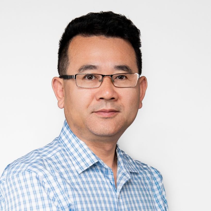
Abstract: Frontotemporal dementia (FTD) is a complex neurodegenerative disorder, involving heterogeneous subtypes such as behavioral variant frontotemporal dementia (BV-FTD), semantic variant frontotemporal dementia (SV-FTD), and progressive non-fluent aphasia frontotemporal dementia (PNFA-FTD). This study aims to inspect connectome-informed changes in brain module organization associated with FTD, identify the potential biomarkers, and gain insights into the pathophysiology for its subtype prediction.
This pilot study illuminates the diverse brain module organization in different FTD subtypes, offering insights into the neurobiological difference that underlies the clinical heterogeneity of FTD. Regions with altered modular properties might also serve as the image markers for predication, early diagnosis, and targeted therapeutic interventions of FTD.

Bio: Prof. Zhen Yuan is a full professor with the Faculty of Health Sciences (FHS)/Centre for Cognitive and Brain Sciences at University of Macau (UM). His research mainly focuses on biomedical optics, cognitive neuroscience, and neuroimaging. Professor Yuan has published over 260 papers in high profile journals in his fields such as Nature Communication, Research, Neuroimage, Cerebral Cortex, Cortex, and Human Brain Mapping. He is the editorial board member of Quantitative Imaging in Medicine and Surgery, associate editor of BMC Medical Imaging, and associate editor of Frontiers in Human Neuroscience.

Prof. Cheng Teng IP

12:47 – 14:15

Lunch at Conference Venue

Keynote Talk

14:15 – 14:50

Abstract: Brain atlas is an indispensable tool for studying the relationship between brain structure and cognitive function. We proposed to create a new brain atlas – the Brainnetome atlas, using brain connectivity profiles. The Brainnetome atlas lays the foundation for research in brain science and brain-inspired intelligence, and opens a new avenue not only for the study of brain science and brain diseases, but also for brain-inspired intelligence. In this lecture, we first introduce the research background and content of the Brainnetome, including the definition and the main research directions of the Brainnetome, the idea of creating the Brainnetome atlas, and the essential differences from existing brain atlas. Then, we will introduce the application of the Brainnetome atlas in elucidating brain cognitive mechanisms and precise diagnosis of brain diseases. Then, we will introduce the challenges and solutions of neuromodulation robots guided by the Brainnetome atlas for precise treatment of brain diseases. Finally, a summary and perspective on future research directions are provided.

Bio: Prof. Tianzi Jiang is Professor and Director of the Brainnetome Center at the Institute of Automation, Chinese Academy of Sciences, and Director of Xiaoxiang Institute of Brain Health. He obtained PhD degree at Zhejiang University and BSc degree at Lanzhou University. He is Chair of Organization of Human Brain Mapping. He was elected a member of the Academy of Europe, a fellow of IEEE, IAPR and AIMBE. He has published over 400 peer-review journal papers and they obtained 40000 citations. He is the recipient of Hermann von Helmholtz Award, Turan Itil Career Contribution Award, and Natural Science Award of China.

Prof. Haoyun ZHANG

14:50 – 15:25

Abstract: Humans rely on safe and secure social environments to thrive as a social species. Research findings indicate that perceived loneliness (loneliness) is a cognitive and mental health risk factor. Our previous work has also revealed that loneliness, when combined with a depressed mood, was negatively correlated to the general cognitive status of older adults. In collaboration with scientists and clinical researchers from Taiwan, the US, and Hong Kong, we conducted a series of studies to understand the neurobiological basis of loneliness in late-life depression (LLD). Our findings indicate that grey matter volumes of the left putamen, caudate, and pallidum could differentiate between healthy controls and people suffering from LLD. Furthermore, a loneliness-related structural sub-network was found across the clinical participants with LLD.

Bio: Prof. Tatia Lee is the Chair Professor of Psychological Science and Clinical Psychology and May Endowed Professor in Neuropsychology at The University of Hong Kong and a visiting Professor at King’s College London. Her research spans the frontiers of neuropsychology and human neuroscience, focusing on the neuroplastic basis of neurocognitive and affective processes underpinning normal and pathological psychological functions. Professor Lee has an impressive publication record of over 300 influential scientific articles. She has achieved significant international and national recognition for her outstanding contributions to the advancement of science. She was elected as a Fellow of esteemed international societies, including The World Academy of Sciences and the UK Academy of Social Sciences.

Prof. Haoyun ZHANG

Main SessionGround Floor

Guest Talks

15:25 – 15:48

Abstract:阿尔茨海默症(AD)特征为记忆与认知障碍,尚无法有效治疗。前驱期pAD是干预关键期,Aβ病理阳性但未达痴呆,诊断干预仍具挑战。多模态磁共振技术精准评估AD脑变,结构成像显示灰质变化,弥散张量呈现微观损伤,新兴QSM、CEST、ASL结合fMRI揭示铁沉积、脑血氧等新视角。聚焦磁声耦合刺激技术,凭借其高空间分辨率和深穿透力的优势,无创调控神经网络,促进递质释放与突触形成,改善脑血流代谢,有望有效缓解AD的病理进程。

Bio: Appointed chairman, Branch of Medical Imaging Engineering and Technology, Chinese Society of Biomedical Engineering; Standing Committee member of the Imaging Branch of the China Medical Rescue Association; Vice President of the Radiological Imaging Equipment Branch of the China Association of Medical Equipment; Vice President of the Imaging Branch of the China Medical Rescue Association; Standing Committee Member of the Radiology Professional Committee of the Chinese Research Hospital Association; Standing Committee Member of the Radiation Health Professional Committee of the China Health Supervision Association; Member of the Branch of Radiation Medicine and Protection of the Chinese Medical Association; Member of the Medical Artificial Intelligence Branch/Intellectual Property and Standardization Working Committee of the Chinese Society of Biomedical Engineering; Member of the expert group for the key projects “Research and Development of Digital Diagnosis and Treatment Equipment” and “Brain Science and Brain-Like Research” under the National Key R&D Program of the Ministry of Science and Technology. Reviewer for the National Natural Science Foundation of China (NSFC). Reviewer for national and Beijing science and technology awards. Honorary Editor of the International Journal of Radiation Medicine; Associate Editor of Artificial Intelligence in Medical Imaging; Editorial Board Member and Reviewer for several journals, including the Chinese Medical Journal, Frontiers in Neuroscience, Frontiers in Oncology, and Brain Imaging and Behavior.

Prof. Haoyun ZHANG

15:48 – 16:11

Abstract: 原子磁力计脑磁图是一种基于2003年发现的全新物理学原理,使用无需低温超导环境的原子磁力计探测脑内神经电活动的新型脑功能成像设备。该技术实现了更高的磁探测灵敏度,不仅进一步提高了脑磁图对脑内电活动信号进行高时空分辨率重建的能力,而且大幅减小了设备的体积,重量和成本,可进行穿戴式记录,并可与VR等设备结合实现头部自由运动状态记录。新型脑磁图的这些特点使其成为了一种在认知科学,心理学,脑机接口,儿童发育心理学,精神类疾病研究,交互心理学等领域极具潜力的新型技术。本次报告介绍该领域的最新进展,技术特点,本实验室的相关工作并和各位老师共同探讨其在各领域的未来应用方向。

Bio: Senior Engineer, Institute of Biophysics, Chinese Academy of Sciences, Director of Magnetoencephalogy Laboratory, State Key Laboratory of Brain and Cognitive Sciences, Beijing Magnetic Resonance Brain Imaging Center, main research interests are magnetoencephalogy equipment, data reconstruction algorithm and its application, presided over the construction, installation, commissioning and operation of the first set of magnetoencephalogy system for scientific research in China. A series of advances have been made in the study of focal localization of UHF concussion epilepsy. The first prototype of a new multi-channel magnetoencephalogram system based on atomic magnetometer was developed in China, and the high-quality magnetoencephalogram signal was successfully obtained, and some leading work was made in the field of cognitive and pre-clinical research of wearable magnetoencephalogram.

Prof. Haoyun ZHANG

16:11 – 16:26

Refreshment & Poster Session

16:26- 16:49

Abstract: Leucine-rich glioma-inactivated protein 1 (LGI1), a secretory protein in the brain, plays a critical role in myelination; dysfunction of this protein leads to hypomyelination and white matter abnormalities (WMAs). Here, we hypothesized that LGI1 may regulate myelination through binding to an unidentified receptor on the membrane of oligodendrocytes (OLs). To search for this hypothetic receptor, we analyzed LGI1 binding proteins through LGI1-3×FLAG affinity chromatography with mouse brain lysates followed by mass spectrometry. An OL-specific membrane protein, the oligodendrocytic myelin paranodal and inner loop protein (OPALIN), was identified. Conditional knockout (cKO) of OPALIN in the OL lineage caused hypomyelination and WMAs, phenocopying LGI1 deficiency in mice. Biochemical analysis revealed the downregulation of Sox10 and Olig2, transcription factors critical for OL differentiation, further confirming the impaired OL maturation in Opalin cKO mice. Moreover, virus-mediated re-expression of OPALIN successfully restored myelination in Opalin cKO mice. In contrast, re-expression of LGI1-unbound OPALIN_K23A/D26A failed to reverse the hypomyelination phenotype. In conclusion, our study demonstrated that OPALIN on the OL membrane serves as an LGI1 receptor, highlighting the importance of the LGI1/OPALIN complex in orchestrating OL differentiation and myelination.

Bio: Prof. Yun Stone Shi, Ph.D, Principal Investigator in Guangdong Institute of Intelligence Science and Technology. Stone obtained his Ph.D degree in physiology at Georgia State University, where he studied vascular ATP-sensitive K channels. He had postdoctoral training in neuroscience with Roger A. Nicoll in UCSF. He started independent research in the Model Animal Research Center at Nanjing University in 2013. From 2022, he joined Guangdong Institute of Intelligence Science and Technology as a senior investigator. His research interest focused on understanding the molecular mechanisms of synaptic function and plasticity, and associated cognitive disorders, if disrupted.

Prof. Haiyan WU

16:49 – 17:12

Abstract: Photoacoustic (PA) imaging is an emerging hybrid imaging technique that can non-invasively identify tissue with high specificity and micron-scale resolution at increased penetration depth. It employs a pulsed laser as the excitation source and gathers the ultrasonic response to reconstruct the maps of light absorption to reflect the structural and functional details of the tissue region. Depending on how exciting light and received sound are aligned, photoacoustic imaging can be multiscale, ranging from human organs and small animal whole body down to microscale fine structures like single cells. The vascular specificity of PA imaging allows for neurovascular coupling of neural voltage imaging, but most works so far interrogate neuronal voltage activities through vascular and blood oxygenation fluctuations rather than direct measurement. Here we propose a novel strategy that employs a whole-field photoacoustic brain detection platform adapted with a photostable voltage sensitive dye to directly monitor voltage dynamics over extended periods in an intact epileptic mouse brain. By investigating the connectivity among brain regions, one can reveal the electrical conduction pathways and their directionality that is indicated through fast temporal visualization. The provided evidence highlights the potential of proposed method for diagnosis and mapping of epilepsy and other voltage-related diseases.
Bio: Dr. Puxiang Lai is a tenured Associate Professor at Department of Biomedical Engineering of the Hong Kong Polytechnic University. His research centers around deep-tissue optical focusing, imaging, stimulation, and treatment. Current research projects include, but are not limited to, wavefront shaping, photoacoustic imaging, neuron imaging, computational optics, and artificial intelligence. His research has fueled more than 100 publications in journals like Nature Photonics, Nature Communications, The Innovation, eLight, Light: Science & Applications, Advanced Photonics, PhotoniX, and Advanced Science. He has been invited to give more than 100 seminars or invited talks worldwide. Dr. Lai was awarded the 2016-2017 Hong Kong RGC Early Career Award.

Prof. Haiyan WU

17:12 – 17:35

Abstract: Cognition typically involves cooperation between multiple brain regions, so thus cognitive states may be detectable by analysis of patterns in functional connectivity between brain regions. We aim to develop a general framework for detecting cognitive states from EEG recordings where we build functional connectivity networks and from them compute various network indices. A highlight of our approach is the analysis of the dynamics in brain region cooperation–how brain network structure changes over short time periods. We apply our methods to the evaluation of mental workload during simulated drone-piloting and detection of mind wandering during video-lecture learning. Our results show that patterns in network dynamics have the potential to discriminate between cognitive states.
Bio: Prof. Zheng Li received a Ph.D. in computer science from Duke University in 2010. After post-doctoral training at Duke in the Nicolelis Lab, he joined the State Key Laboratory of Cognitive Neuroscience and Learning at Beijing Normal University in 2013. In 2020 he moved to the State Key Lab’s Center for Cognition and Neuroergonomics at Beijing Normal University at Zhuhai. His research interest is in brain-computer interfaces, focusing on methods for processing and decoding neural activity, with recent work mainly using non-invasive electroencephalograph recordings.

Prof. Haiyan WU

Parallel Session – 1st Floor 1005

Guest Talks

15:30 – 15:53

Abstract: The central focus of cognitive neuroscience lies in elucidating the relationship between brain activity and information processing. There is substantial empirical evidence indicating that the brain processes various types of cognitive inputs, such as speech and language, in a predictive manner. The brain activation elicited by stimulation often represents only a small fraction of the energy expenditure associated with the resting-state predictive activity. Consequently, brain activation can be conceptualized as the reorganization of activity flows relative to their organization at baseline. These dynamic changes can be estimated by calculating the local differences in activity between adjacent voxels, i.e., gradients of activity, which have been shown to be stimulus-specific. Furthermore, the differences between adjacent voxels form spatial patterns of activity that are consistent across brain regions involved in the relevant functional networks. Thus, fMRI reflects the spatial coding of the processed information, which can complement the temporal coding information obtained from electrophysiological measurements. The integration of neuroimaging techniques and psychophysical approaches has the potential to enhance our understanding of phenomena such as addiction, decision-making, and language.

Bio: Prof. Strelnikov graduated from the Faculty of Medicine of Saint-Petersburg University, where also defended his PhD thesis in Physiology and Neuroscience. Afterwards, he did several postdocs at the Brain and Cognition Center of Helsinki University, the Center of cognitive neuroimaging of Paris University, and the Brain and Cognition Center of Toulouse University. Then he worked as clinical research coordinator and chief of research projects at the Direction of clinical research of the Toulouse University Hospital Center. From November 2023, he worked as associate professor in Center of Cognitive and Brain Sciences, University of Macau.

Prof. Michiel SPAPÉ

15:53 – 16:16

Abstract: Aging is often accompanied by declined cognitive function, leading to a high risk of dementia. In particular, older adults often have difficulty in processing complex information such as making decisions under risk. Our recent researches have reported that the fronto-subcortical network plays a crucial role in age-related cognitive deterioration, and could be a possible interventional target. In risky decision-making tasks, we found close relationships between fronto-subcortical networks and risk-taking behaviors in older individuals. Using transcranial electric stimulation, the frontal stimulation shows beneficial effects on brain efficiency in multiple domains, such as improved working memory, better decision strategy and less cognitive fatigue. In rodent models, we also found that TMS with extremely low intensity remarkably modulated cognitive performances, depending on stimulation frequency and intensity. Although non-invasive brain stimulation could be an effective interventional approach, more research is needed to optimize experimental design and parameters.

Bio: Prof. Ren Ping received PhD degree (Neurobiology) from Chinese Academy of Sciences, and continued postdoc research in University of Rochester for 6 years. Since 2019, he joined Shenzhen Mental Health Center/Shenzhen Kangning Hospital. His research focuses on the neural substrates of cognitive aging and dementia, and early intervention of cognitive impairment. He uses multiple approaches, including computerized behaviour tasks, non-invasive brain stimulation, and MRI. His recent work is mainly about decision-making alterations and early intervention in older adults.

Prof. Michiel SPAPÉ

16:16 – 16:26

Refreshment & Poster Session (Ground Floor)

16:26 – 16:49
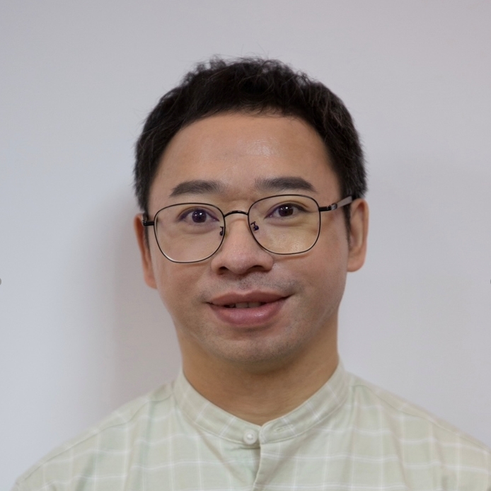
Abstract: Investigation into early embryonic functions, e.g. brain activity, have long been constrained due to the technical challenges involved. Functional ultrasound (fUS) has emerged as a breakthrough modality for real-time monitoring of brain activity, offering considerable potential as a tool for studying functional embryonic development. In the present study, fUS was leveraged to monitor the functions of developing mice from embryonic days E8.5 to E18.5, revealing its ability to capture whole-embryo activity with exceptional spatial and temporal resolutions. The data revealed a high correlation between cardiac function and body size, underscoring the pivotal role of cardiac function in embryonic growth. Moreover, brain activity across the gestational period was successfully captured, providing valuable information about brain activity during embryonic/fetal development. Thus, our study offers novel insights into embryonic functional development, laying the foundation for embryonic fUS imaging in both scientific research and clinical contexts.
Bio: Prof. Zhihai Qiu received his B.S. from Fujian Normal University in August 2012. He received his Ph.D. of Biomedical Engineering from The Hong Kong Polytechnic University, Hong Kong in April 2019. Afterwards He obtained postdoctoral training at Stanford and became a principal investigator at Guangdong Institute of Intelligence Science and Technology consequently, working on multiscale brain imaging and neuromodulation technologies.

Prof. Rihui LI

16:49 – 17:12

Abstract: Cerebral small vessel disease (CSVD) is the most prevalent cerebrovascular condition among the elderly. It is one of the leading causes of disability, functional loss, and cognitive decline in this population. The diagnosis of CSVD relies on the Standards for Reporting Vascular Changes on Neuroimaging (STRIVE-II). However, traditional MRI techniques are limited by signal-to-noise ratio and contrast, making it difficult to detect early microvascular changes.
With the advent of ultra-high field (UHF) MRI, high-resolution neurovascular imaging at 7 Tesla (7T) can clearly depict the structures and lesions of cerebral small vessels, particularly the lenticulostriate arteries and deep medullary veins. In this presentation, we will demonstrate the effectiveness of accelerated high-resolution imaging sequences at 7T for these arterioles and venules. Meanwhile, we will introduce our neural network-based algorithms for small vessel segmentation and modeling. Utilizing these advanced UHF-MRI methods, we have identified various pathological features of cerebral small vessels in CSVD and revealed neurovascular imaging markers most directly associated with cognitive and behavioral decline.
UHF neurovascular imaging provides significant insights into the pathophysiology of CSVD, offering potentials in understanding its pathogenic mechanisms on brain cognition and function, as well as in improving the diagnosis and management of CSVD.
Bio: Prof. Zhang Zihao is the manager of the 7T MRI scanner at the Institute of Biophysics, Chinese Academy of Sciences (CAS). He is also overseeing the construction of the 10.5T human scanner at the Institute of Artificial Intelligence, Hefei Comprehensive National Science Center. Prof. Zhang’s research focuses on developing ultra-high field neuroimaging techniques, particularly for neurovascular imaging. He has identified various imaging markers of cerebral small vessel lesions. He is a member of the Youth Innovation Promotion Association of the CAS and was supported by the Young Elite Scientists Sponsorship Program of the China Association for Science and Technology.

Prof. Rihui LI

17:12 – 17:35

Abstract: Brain-computer interfacing enable people to interact with the outside world by classifying motor imagery while EEG classification has shown ability to detect and visualize perception. Can neuroimaging allow us to determine what somebody is thinking of? To get to this promised centre of cognition, I argue more is required than merely matching perception or action with patterns of neural activity. Instead, to gain insight into consciousness an understanding of the three-directional relations between stimuli, cognition and brain activity is required. I introduce the neuroadaptive interface as a technique specifically designed to model these relations: By adapting a generative neural network to neural activity related to cognitive processes such as categorisation or attention, a model learns to associate human, subjective dimensions to its latent representation. Furthermore, the use of generative artificial intelligence enables us to provide on-line visualization related to contents of consciousness. Previous work in this area has demonstrated accurate recreation of individual categorisation of faces on the basis of subjective qualities such as age, emotion, or attractiveness. In the present talk, I will demonstrate how the research programme has developed to now provide models of memory, visualizing facial memories and their relation to short-term recognition and long-term recall.
Bio: Prof. Michiel Spapé received his PhD in Psychology from Leiden University (2009). After several postdoctoral positions in England (University of Nottingham), Finland (Aalto / Helsinki University), and a brief time in Liverpool as Assistant Professor, he was invited to join the University of Macau as Associate Professor. Here, he studies cognition, consciousness and emotion using a combination of neuroimaging (EEG) and digital (AI, VR) technologies. He has published over 70 articles (cited ca. 2150), two textbooks on experimental design and neuroimaging, and a patent on information retrieval using brain-computer interfacing.

Prof. Rihui LI

17:35 – 17:45

  Break

17:45 – 18:05

  Poster Awards

   Closing Remarks & Group Photos

18:20

   Dinner for Invited Speakers at Grand Plaza 萬豪軒 (N1 Ground Floor)